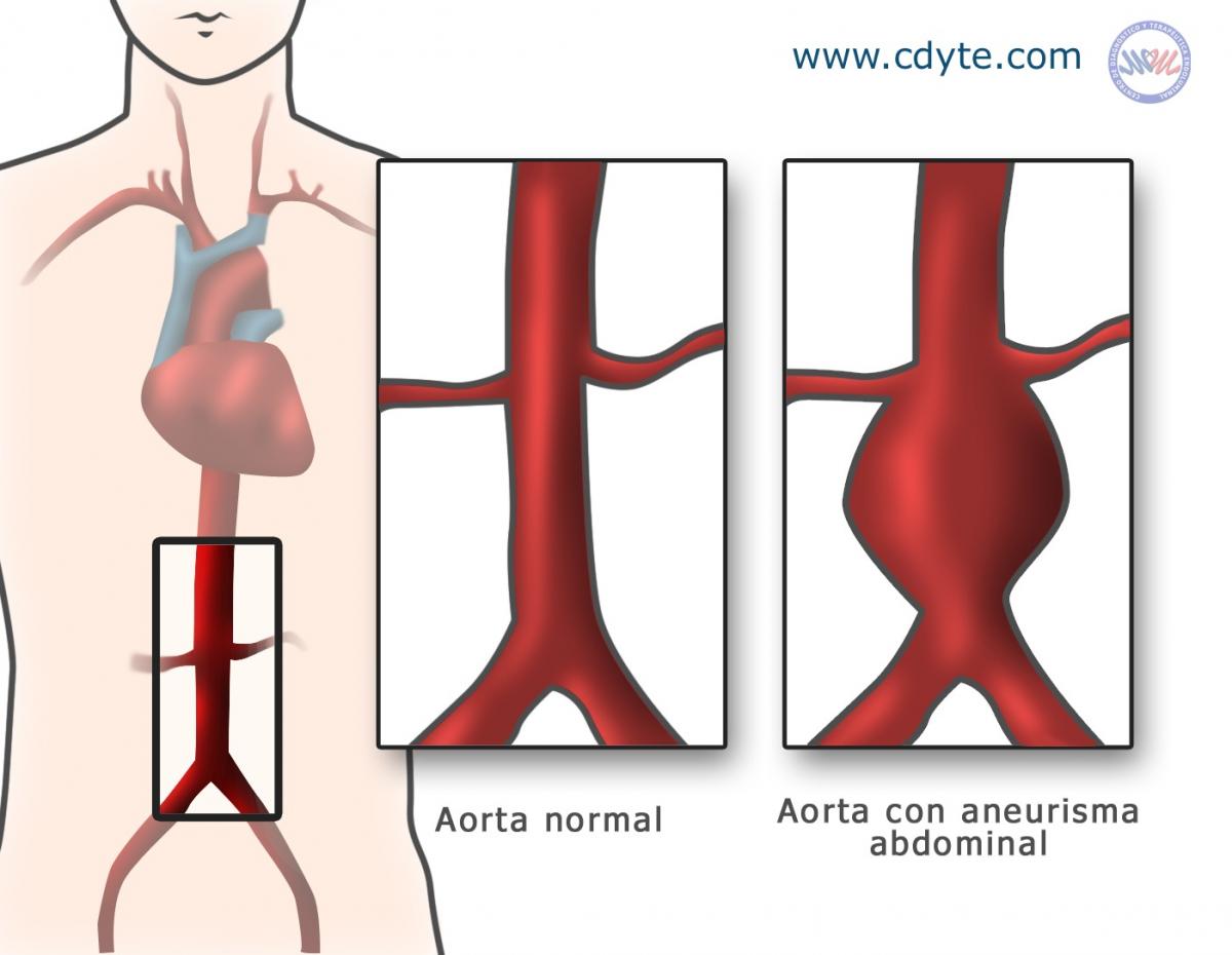
What is an Abdominal Aortic Aneurysm? 
An Abdominal Aortic Aneurysm (AAA) consists of a dilation of the aorta, which is the most important blood vessel in the body, which comes out of the heart and branches off to all the organs. Aneurysms may appear in any of the blood vessels in the human body, although the most common is the abdominal aorta, below the renal arteries (the blood vessels supplying the kidneys). An aneurysm can grow until, like a balloon, it bursts. The bigger the aneurysm, the easier is grows. The rupture of an aneurysm can be fatal. The aim of all surgical procedures on aneurysms is to avoid rupture.
How common are they?
Aneurysms are four times more common in men than in women and, generally, appear after 55-60 years of age. As the population ages the incidence of aortic aneurysms increases every ten years.
Who is at risk of suffering AAA?
Those most at risk are:
- Men, the disease is 4 times more common in men than in women.
- Those over 60 years of age.
- Smokers.
- Those whose family has a history of abdominal aneurysm or in any other part of the body.
- Arteriosclerosis (hardening of the arteries).
- Those with high blood pressure.
- Patients with heart disease.
- Those whose family has a history of vascular disease.
What are the symptoms of AAA?
An Abdominal Aortic Aneurysm ruptures in most cases with no warning. The condition, which affects both men and women, does not generally present symptoms. When there are symptoms, they are mainly:
- Intense abdominal pain, which may or may not be constant.
- Lower back pain, which may be reflected in other parts of the body.
- A throbbing sensation in the abdomen.
- Weakness.
- On occasions the lump in the abdomen can be felt when pressing the area.
The rupture of an aneurysm is very serious and the main symptoms are:
- Sudden, intense pain.
- Paleness
- Rapid pulse.
- Dry mouth and thirst.
- Nausea and vomiting.
- Fainting
- Profuse perspiration.
- Shock.
In the case of suspected rupture, you must see a doctor immediately.
How is an AAA diagnosed?
Apart from a clinical examination that guides the diagnosis, image diagnosis methods can be used such as: Ultrasound, Computerized Tomography, Magnetic Resonance and Arteriography.
How can AAA be treated?
If the aneurysm is small, it is controlled using image diagnosis methods. If it reaches a certain size or is growing rapidly, surgery may be required. Normally, the aorta measures about 2.3 cm in diameter in men and 1.9 cm in women. Aneurysms measuring 5 cm or more in diameter require surgery. There are two possible treatments:
- Open surgery. The surgeon makes an incision in the abdomen and inserts a tube made of a special material.
- Endovascular treatment (minimally invasive technique). Before the procedure, the doctor analyzes the image studies carried out beforehand (CT scans, angiograms). The doctor can then choose the right stent graft for each patient. The doctor makes small incisions in each groin to access the femoral arteries (blood vessels in the legs).
Guided by images on an x-ray machine, the doctor, through the arteries in the groin, inserts a catheter containing a stent and guides it to the aorta. The stent graft is covered with a special synthetic material and the catheter has a device with which to release the stent. When it is correctly positioned, the stent graft is released, expanding to the right diameter and preventing blood from reaching the aneurysm, the catheter is then removed. The bottom of the stent graft is divided in two and, in some cases one or two extensions (a stent of a smaller caliber than the principal stent) are put in place depending on the model used. Thus, blood flow through the aorta, the pelvic arteries and legs continues without filling the aneurysm (closure or exclusion of the aneurysm).
[video:http://www.youtube.com/watch?v=yVZYKsdsNiU]